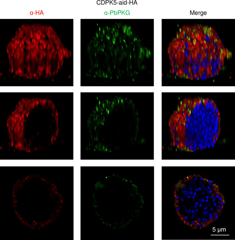
Fig 2. CDPK5 and PKG are present in the periphery of merosomes.

Representative deconvolved images and optical sections of immunostained CDPK5-aid-HA merosomes. The top panel illustrates a volumetric view of a merosome. Middle and bottom panels illustrate longitudinal optical sections of the merosome.
