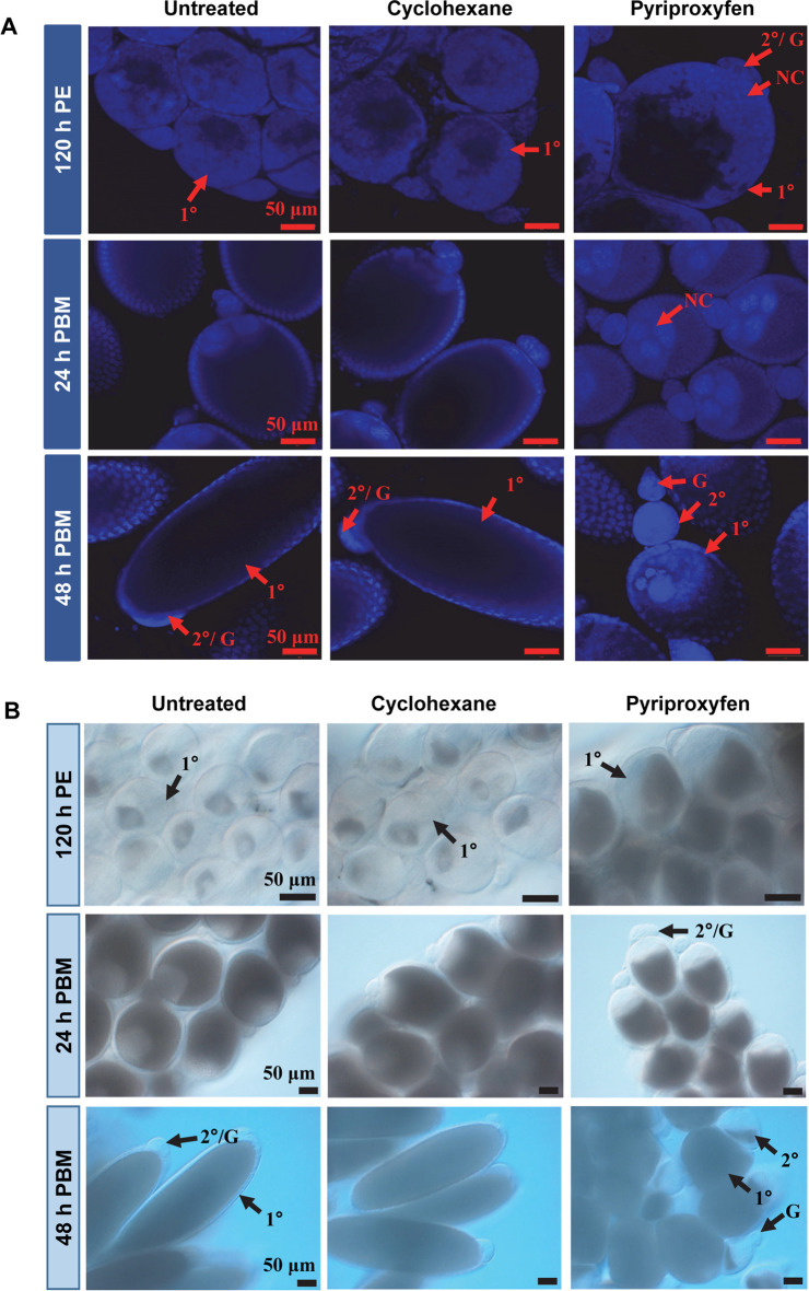
Fig 1. Follicular development in PPF-treated mosquitoes.
(A) Ovarian follicles were collected at the indicated time points after PPF treatment. After fixation, follicles were stained with DAPI. Images were captured using a Zeiss LSM 880 confocal microscope. Scale bars represent 50 μm. (B) Differential interference contrast (DIC) images of ovarian follicles after PPF treatment. PE, Post eclosion; PBM, Post blood-meal. 1°, primary follicle; 2°, secondary follicle; G, germarium; NC, nurse cell.

