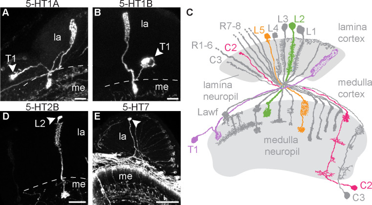
Fig 1. Lamina neurons including T1 and L2 express serotonin receptors.
(A-E) Serotonin receptor MiMIC-T2A-GAL4 lines were crossed to UAS-MCFO-1 to sparsely label individual cells in the lamina. Cell bodies are indicated by an arrowhead. 5-HT1A (A) and 5-HT1B (B) MCFO crosses revealed cells with morphologies identical to T1 neurons. (C) A diagram showing lamina neurons adapted from [80] highlights L2 (green), L5 (orange), T1 (purple), and C2 (pink). (D) 5-HT2B>MCFO labeled cells were morphologically identical to L2 neurons. (E) 5-HT7>MCFO-1 labeled neurons possibly representing L5 lamina monopolar cells. Scale bars are 20 μm and N = 9–31 brains imaged per receptor subtype. Due to the nature of stochastic labeling, some cell types were observed in only a subset of brains: 22/31 (A), 10/11 (B), 9/9 (D), and 7/13 (E).

