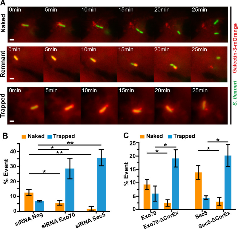
Fig 4. The exocyst plays a role in the displacement of BCV membrane remnants from S. flexneri.
(A) Time-lapse microscopic acquisition of the fate of S. flexneri BCV was analyzed. Images were recorded every minute and the z-projections of representative infection foci were shown. S. flexneri expressing eGFP was employed (green), while ruptured BCV was indicated by Galectin-3-mOrange (red). Scale bars are 2 μm. Representative images of S. flexneri moving free of BCV membrane (“Naked”, upper panel), S. flexneri moving with some BCV remnants (“Remnant”, middle panel) and S. flexneri moving with the damaged vacuole (“Trapped”, bottom panel). Analysis of the fates of individual S. flexneri and their BCVs with reference to the observations in (A) in different conditions including (B) RNA interference of non-targeting control (siRNA Neg) versus Exo70 depletion (siRNA Exo70) or Sec5 depletion (siRNA Sec5) and (C) expression of Exo70 or Sec5 versus their mutants lacking the core exocyst assembly motif (Exo70-ΔCorEx and Sec5-ΔCorEx, respectively). Data are shown as mean ± SEM in triplicates of each condition (*p<0.05; **p<0.01).

