. 2020 Apr 20;1(1):32–47. doi: 10.1158/2643-3230.BCD-19-0028

Figure 4.

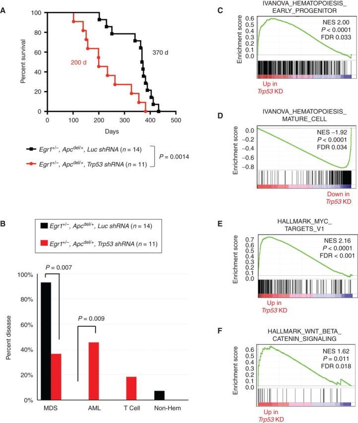
Figure 4. Following exposure to an alkylating agent, haploinsufficiency of both Egr1 and Apc promotes development of MDS; loss of these genes together with Trp53 promotes AML development. A, Kaplan–Meier survival curves of WT recipients transplanted with Egr1+/−, Apcdel/+ BM cells transduced with luc shRNA (EA-luc: black) or Trp53 shRNA (EA-Trp53: red). Both donor and recipient mice were treated with ENU. Disease development is significantly faster in EA-Trp53 mice compared with EA-luc mice (200 days vs. 370 days, P = 0.0014). B, Histologic classification of diseases shows that most EA-luc mice developed MDS, and none developed AML. C–F, Mouse genes were collapsed to human gene names; differentially expressed genes in EA-Trp53 AML samples (n = 6) versus EA-luc MDS samples (n = 6) were analyzed using the GSEA software. Features, such as a block in myeloid differentiation and upregulation of MYC target genes and WNT/β-catenin signaling recapitulate human t-MN.

Following exposure to an alkylating agent, haploinsufficiency of both Egr1 and Apc promotes development of MDS; loss of these genes together with Trp53 promotes AML development. A, Kaplan–Meier survival curves of WT recipients transplanted with Egr1+/−, Apcdel/+ BM cells transduced with luc shRNA (EA-luc: black) or Trp53 shRNA (EA-Trp53: red). Both donor and recipient mice were treated with ENU. Disease development is significantly faster in EA-Trp53 mice compared with EA-luc mice (200 days vs. 370 days, P = 0.0014). B, Histologic classification of diseases shows that most EA-luc mice developed MDS, and none developed AML. C–F, Mouse genes were collapsed to human gene names; differentially expressed genes in EA-Trp53 AML samples (n = 6) versus EA-luc MDS samples (n = 6) were analyzed using the GSEA software. Features, such as a block in myeloid differentiation and upregulation of MYC target genes and WNT/β-catenin signaling recapitulate human t-MN.