
Fig. 6. Phosphorylation of ubiquitin mediated DNA repair blockage in cells at low stiffness.
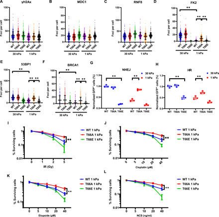
(A to F) Ubiquitin-replacement HEK293 cells expressing wild-type (WT), T66A, or T66E mutant ubiquitin were plated on soft (1 kPa) and stiff (30 kPa) fibronectin-coated hydrogels. Cells were fixed 1 hour after irradiation and stained with anti–γ-H2AX (A), MDC1 (B), RNF8 (C), FK2 (D), 53BP1 (E), and BRCA1 (F) antibodies. Data are presented as means ± SD, n = 3 biologically independent samples (**P < 0.01). (G and H) Phosphorylation of ubiquitin inhibits HR and NHEJ. Effect of ubiquitin phosphorylation on the efficiency of NHEJ (G) and HR (H) was analyzed by flow cytometry. Data are presented as means ± SD. n = 3 biologically independent samples (**P < 0.01). (I to L) Phosphorylation of ubiquitin regulates genotoxic sensitivity. Ubiquitin-replacement HEK293 cells expressing wild-type (WT), T66A, or T66E mutant ubiquitin were plated on soft (1 kPa) fibronectin-coated hydrogels. Cells were treated with indicated genotoxic agents. Colony formation assays were performed to examine survival of cells expressing wild-type (WT), T66A, or T66E mutant ubiquitin on soft (1 kPa) fibronectin-coated hydrogels. Data are presented as means ± SD. n = 3 biologically independent samples.