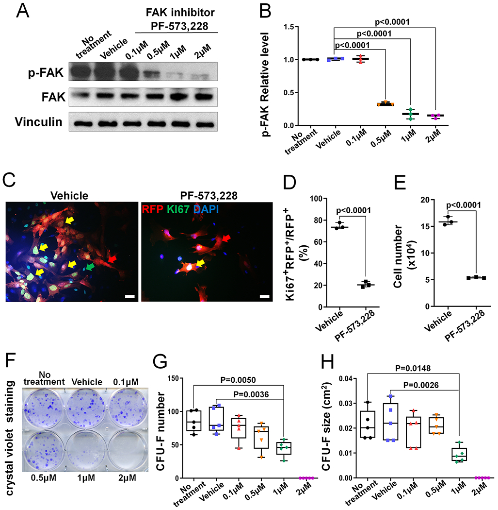
Figure 4. FAK kinase inhibition leads to decreased BMSC proliferation.

(A) Representative western blot image showing that phospho-FAK (p-FAK) level in BMSCs was dose-dependently suppressed by FAK kinase inhibitor PF-573,228. (B) Quantification of the p-FAK relative level in controls (no treatment and vehicle treatment) and FAK inhibitor-treated BMSCs. p-FAK was normalized to total FAK protein (n=3). (C) Representative fluorescent images of BMSCs isolated from CHet-RFP mice after the treatment with vehicle or FAK inhibitor PF-573,228 for 7 days. Yellow arrows indicate the RFP+Ki67+DAPI+cells; red arrows indicate the RFP+Ki67− DAPI+cells; green arrows indicate the RFP−Ki67+DAPI+cells. Scale bar=20 μm. (D) Quantification of the percentage of Ki67 and RFP double positive cells in the cultures described in (C) (n=3). (E) Quantification of the total cell number in the cultures described in (C) (n=3). (F) Representative image of crystal violet staining in BMSC culture treated with indicated doses of FAK inhibitor PF-573,228 for 10 days. (G) Quantification of the CFU-F number; and (H) Quantification of the CFU-F size in cultures described in (F). Each data point represents the average of one independent experiment (n=5). Values were presented as median and interquartile range.
