
Extended Figure 11: Restoration of complex II by ectopic expression of SDHA cDNA rescues tumor growth.
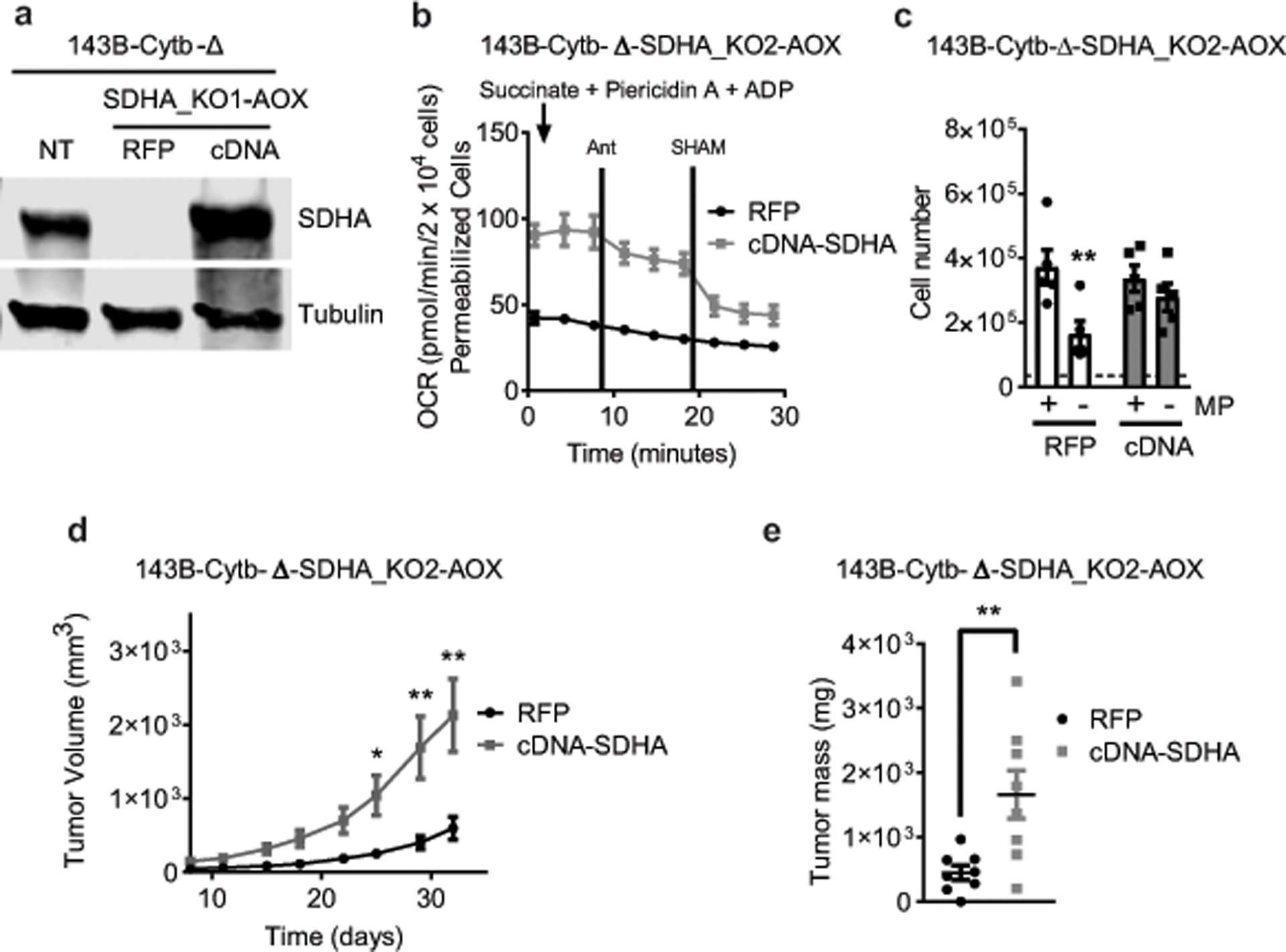
a, Western blot analysis of SDHA protein levels in 143B-Cytb-Δ-NT, 143B-Cytb-Δ-SDHA_KO2-AOX-RFP and 143B-Cytb-Δ-SDHA_KO2-AOX-cDNA SDHA cells. Data representative of three independent experiments. b, Complex II driven oxygen consumption rate of permeabilized 143B-Cytb-Δ-SDHA_KO2-AOX-RFP and 143B-Cytb-Δ-SDHA_KO2-AOX-cDNA SDHA cells. Succinate and ADP were provided as substrates. Piericidin A (1 μM) and Antimycin A (1 μM) were used to inhibit complex I and III respectively. SHAM (2 mM) was used to inhibit AOX activity (n=4 biologically independent experiments). c, 143B-Cytb-Δ-SDHA_KO2-AOX-RFP and 143B-Cytb-Δ-SDHA_KO2-AOX-cDNA SDHA cells were grown in the presence or absence of methyl pyruvate and cell number was assessed after 72h (n=5 biologically independent experiments). d,e, Average tumor volume (d) and tumor mass (e) of xenografts from 143B-Cytb-Δ-SDHA_KO2-AOX-RFP and 143B-Cytb-Δ-SDHA_KO2-AOX-cDNA SDHA cells (n=8 mice per group from two independent cohorts). Data represent mean ± s.e.m. (b-e). Statistical significance was determined using two-tailed t-tests (e) or 2-way ANOVA (c,d) with a Bonferroni test for multiple comparisons (*p < 0.05; **p < 0.01, exact P values in Source Data). For gel source data, see Supplemental Figure 6.