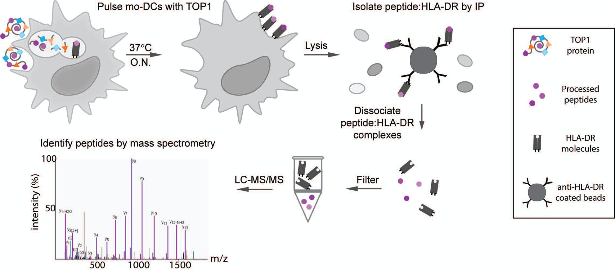
Figure 1. Natural Antigen Processing Assay (NAPA).

MoDCs from 6 donors with ATA-positive scleroderma were incubated with whole TOP1 protein overnight (O.N.) at 37°C. Cells were then lysed and the peptide:HLA-DR complexes isolated by immunoprecipitation using magnetic beads coated with anti-HLA-DR antibodies (clone L243). The presented peptides were dissociated from the HLA-DR molecules using 0.1% TFA and separated from HLA-DR molecules using a 10kDa spin filter, which allows the smaller molecular weight peptides to pass through. The filtered peptides were then identified and sequenced by liquid chromatography with tandem mass spectrometry (LC-MS/MS).
