
Figure 3. Changes to the lungs of rhesus macaques infected with SARS-CoV-2 and treated with remdesivir.
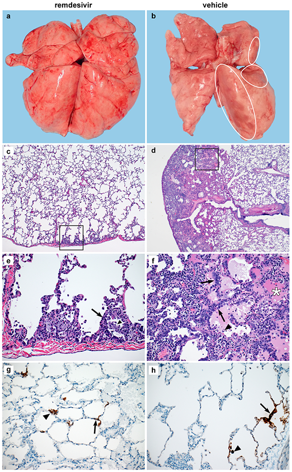
Rhesus macaques infected with SARS-CoV-2 and treated with remdesivir (n=6) or vehicle solution (n=6) were euthanized on 7 dpi. (a) Representative dorsal view of lungs of a remdesivir-treated animal. (b) Representative dorsal view of lungs of a vehicle-treated animal with focally extensive areas of consolidation (circles). Histological analysis was performed on 3 sections from 6 lung lobes from each of the 6 animals per treatment group and representative images were chosen for panels c-h. (c) Minimal subpleural interstitial pneumonia (box) observed in 3 of 6 remdesivir-treated animals. (d) Moderate subpleural interstitial pneumonia with edema (box) observed in 5 of 6 vehicle-treated animals. (e) Boxed area from panel c with alveoli lined by type II pneumocytes (arrow) and alveolar spaces containing foamy macrophages (arrowhead). (f) Boxed area from panel d with pulmonary interstitium expanded by edema and moderate numbers of macrophages and neutrophils. Alveoli are lined by type II pneumocytes (arrows). Alveolar spaces are filled with edema (asterisk) and small numbers of pulmonary macrophages (arrowhead). (g) Viral antigen in type I pneumocytes (arrow) and type II pneumocytes (arrowhead) of a remdesivir-treated animal. (h) Viral antigen in type I pneumocytes (arrow) and macrophage (arrowhead) of a vehicle-treated animal. Magnification c and d: 40x; panel e-h: 200x.