
Figure 3.
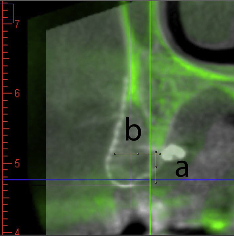
Superimposed images of pre-operative and post-operative CT. The pre-operative CT image was transformed into green shade and matched with the basal bone of the post-operative CT image. The measurement was set as the implant orientation and perpendicular to the occlusal plane. The vertical bone gain (a) was measured from the most coronal part of the original bone contour to the most coronal part of the new bone, and the horizontal bone gain (b) was measured at the widest part of the new bone formation. The horizontal and vertical bone gain results were recorded in Table 1.