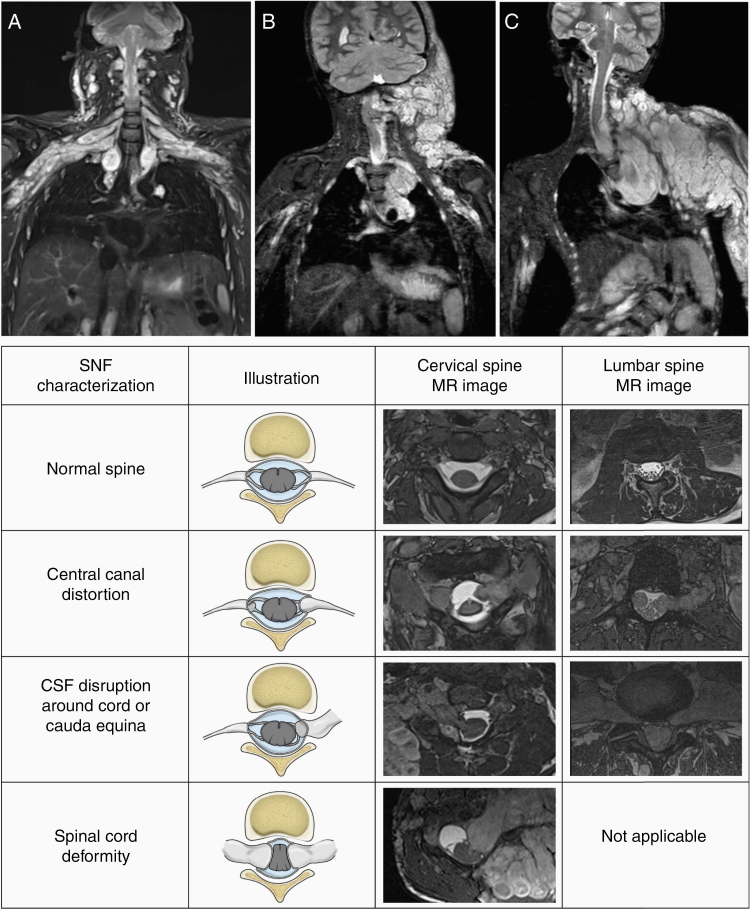
Figure 1.
Spinal neurofibroma (SNF) burden characterization. Coronal short T1 inversion recovery MR images demonstrate the SNF distribution: multilevel symmetrical (A), multilevel predominantly one-sided (B), or single spinal nerve root (C). In addition to SNF, extensive plexiform neurofibroma (PN) is seen in the same body region in all 3 patients. The chart below illustrates our method of capturing the presence or absence of SNF-related spinal canal distortion, cerebrospinal fluid (CSF) disruption around the cord or cauda equina, and spinal cord deformity.

