Figure 2.
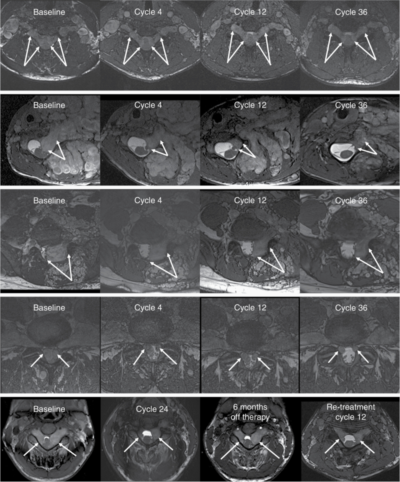
MRI examples of subtle (top row) and marked improvement (rows 2–5) in patients with spinal neurofibroma (SNF) receiving selumetinib therapy. The area of the spinal canal is shown on axial balanced fast field echo sequences. Arrows indicate the SNFs of interest. First row: Bilateral dumbbell tumors compress the spinal cord at the cervical 5–6 level. There is a slight gradual improvement in the narrow wedge shape of the cord and an increase in cerebrospinal fluid (CSF) abundance. Also note the decreasing size of the adjacent brachial plexus plexiform neurofibroma (PN). Second row: SNF in the left cervical 6–7 neuroforamen extends into the central canal and deforms the spinal cord. The size of the mass is markedly reduced after 4 cycles of selumetinib therapy with further improvement through cycle 36. Third row: SNF below the level of the cord (L5-S1) displacing the thecal sac and nerve roots. SNF shrinkage is apparent by cycle 4 and the response is maintained through cycle 36. Fourth row: Lumbar 4–5 level bilateral SNFs completely fill the central canal at baseline. CSF becomes detectable around the nerve roots after 4 treatment cycles and the improvement continues through cycle 30. Fifth row: The spinal cord deformity at C3-4 level is reduced during 24 cycles of therapy. Interruption of selumetinib treatment for 6 months resulted in SNF regrowth; however, improvement is evident again upon retreatment with selumetinib.

