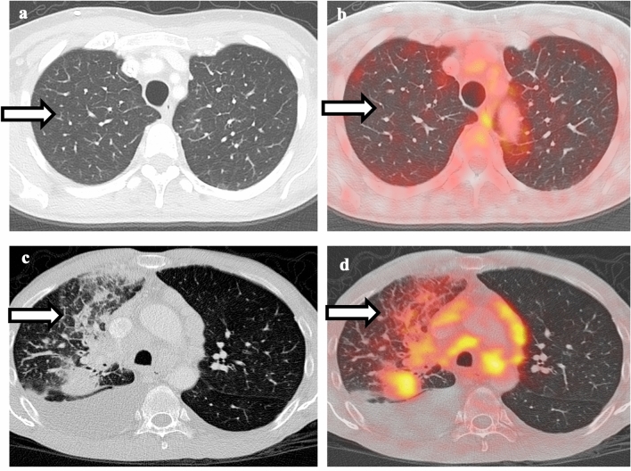
Fig. 2.
Comparison between pulmonary edema and lymphangitic spread of tumor. a Non-contrast CT of the chest shows smooth interlobular septal thickening in bilateral upper lobes. b Fusion of PET with CT shows no associated hypermetabolism, compatible with pulmonary edema. c Non-contrast CT of the chest shows right lower lobe mass, irregular and nodular thickening of interlobular septa, and small right pleural effusion. d Fusion of PET with CT shows hypermetabolism associated with the right lower lobe mass as well as the interlobular septa, compatible with lymphangitic spread of tumor

