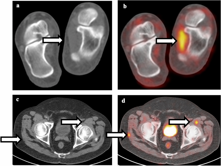
Fig. 9.
Comparison between muscle strain and intramuscular metastatic disease. a Non-contrast CT of the foot shows normal appearance of the left abductor hallucis muscle. b Fusion of PET and CT shows linear hypermetabolism associated with the left abductor hallucis muscle, compatible with muscle strain. c Non-contrast CT of the pelvis shows subtle lesions in the right gluteus maximum and left iliopsoas muscle. d Fusion of PET and CT shows focal intense FDG uptake associated with these lesions, compatible with intramuscular metastatic disease

