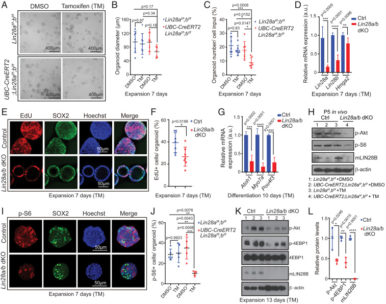
Fig. 6.
Loss of LIN28A/B attenuates mTOR signaling and limits supporting cell proliferation and hair cell formation in cochlear organoid culture. Cochlear organoid cultures were established from UBC-CreERT2;Lin28af/f;Lin28bf/f mice and Lin28af/f;Lin28bf/f littermates’ stage P2. Cultures received 4-hydroxy-tamoxifen (TM) or vehicle control DMSO at plating. SOX2 (green) marks supporting cells/prosensory cells, Hoechst (blue) staining marks cell nuclei. (A–G) Loss of Lin28a/b inhibits cell proliferation and hair cell production in organoid culture. (A) Representative BF images of control organoids (TM: Lin28af/f;Lin28bf/f; DMSO: UBC-CreERT2;Lin28af/f;Lin28bf/f; DMSO: Lin28af/f;Lin28bf/f) and Lin28a/b dKO organoids (TM: UBC-CreERT2;Lin28af/f;Lin28bf/f) at 7 d of expansion. (B) Diameter of control and Lin28a/b dKO organoids in A (n = 7, two independent experiments). (C) Organoid forming efficiency in control and Lin28a/b dKO cultures (n = 7, two independent experiments). (D) qRT-PCR analyzing Lin28a, Lin28b and Hmga2 mRNA expression in Lin28a/b dKO organoids (red bar) compared to control organoids (blue bar) at 7 d of expansion (n = 5 for control, n = 6 for Lin28a/b dKO, two independent experiments). (E) Cell proliferation in control and Lin28a/b dKO organoids. An EdU pulse was given at 7 d of expansion and EdU incorporation (red) was analyzed 1 h later. (F) EdU incorporation in E (n = 7, two independent experiments). (G) qRT-PCR analysis of Atoh1, Myo7a, and Pou4f3 mRNA expression in Lin28a/b dKO organoids (red bar) compared to control organoids (blue bar) at 10 d of differentiation (n = 4, two independent experiments). (H) Immunoblots for LIN28B, p-Akt, p-S6, and β-actin using protein lysates of acutely isolated control and Lin28a/b dKO cochlear epithelia, stage P5. (I–L) Loss of Lin28a/b attenuates mTOR signaling in cochlear organoids. (I) Immunostaining for p-S6 protein (red) in control and Lin28a/b dKO organoids at 7 d of expansion. (J) Percentage of p-S6+ cells in I (n = 5, two independent experiments). (K) Immunoblots for p-Akt, p-4EBP1, 4EBP1, mLIN28B, and β-actin (loading control) using protein lysates of control and Lin28a/b dKO organoids after 7 d of expansion. (L) Normalized p-Akt, p-4EBP1, and m-LIN28B protein levels in control and Lin28a/b dKO organoids in K (n = 3, from one representative experiment, two independent experiments). Bars in D and G represent mean ± SD, otherwise individual data points and their mean ± SD were plotted. Individual data points in B, F, and J represent the average values per animal. n = animals analyzed per group. Two-way ANOVA with Tukey’s correction was used to calculate P values in B, C, and J. Otherwise, P values were calculated using two-tailed, unpaired Student’s t tests.

