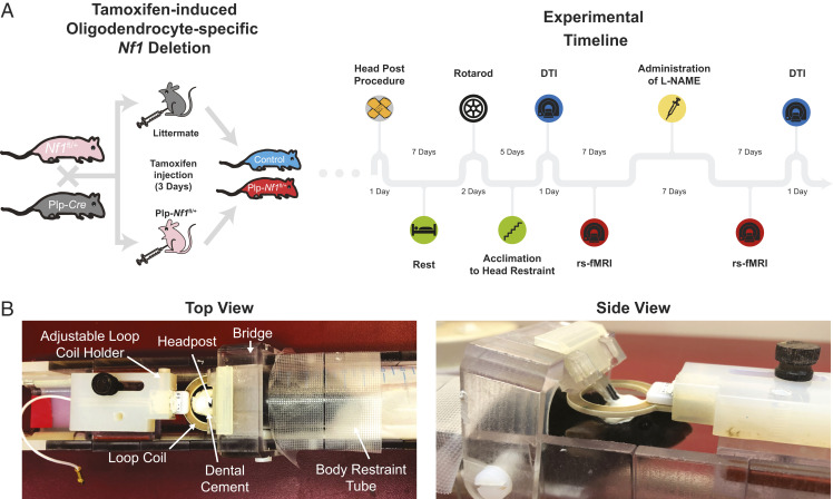
Fig. 1.
Experimental setup for whole-brain imaging of Plp-Nf1fl/+ mice. (A) Oligodendrocyte-specific Nf1 deletion was achieved by crossing of Plp-Cre and Nf1fl/+ mice. Plp-Nf1fl/+ and littermate animals received a 3-d tamoxifen treatment (twice daily) to induce the deletion in Plp-Nf1fl/+ mice and create littermate controls (Plp-Nf1+/+). The experimental timeline is shown for mice that underwent full evaluation including behavior, as well as structural and functional imaging. (B) A custom-built 3D-printed modular cradle allows head fixation for functional scans of awake passive mice. The same cradle allows the addition of a mouse-compatible mask that can be placed around the animal’s nose and mouth to maintain constant anesthesia of the head-fixed mice for long structural scans.

