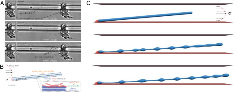
Fig. 1.
In vitro experiments and numerical simulations of platelet biogenesis by Bächer et al. (3). (A) Experimental images of a megakaryocyte growing in vitro and forming a long tubular extension (proplatelet) inside a microchannel. Swellings are seen to form along the tube and will give rise to fragmentation into platelets. (B) Schematic of the computational model, where the proplatelet is represented as a cylindrical membrane tube anchored at a wall and subject to a pressure-driven parabolic flow. (C) Snapshots from a simulation, where swellings are seen to develop along the membrane tube by an active Rayleigh–Plateau instability and will ultimately break up into platelets. Adapted from ref. 3 with permission.

