
Figure 1.
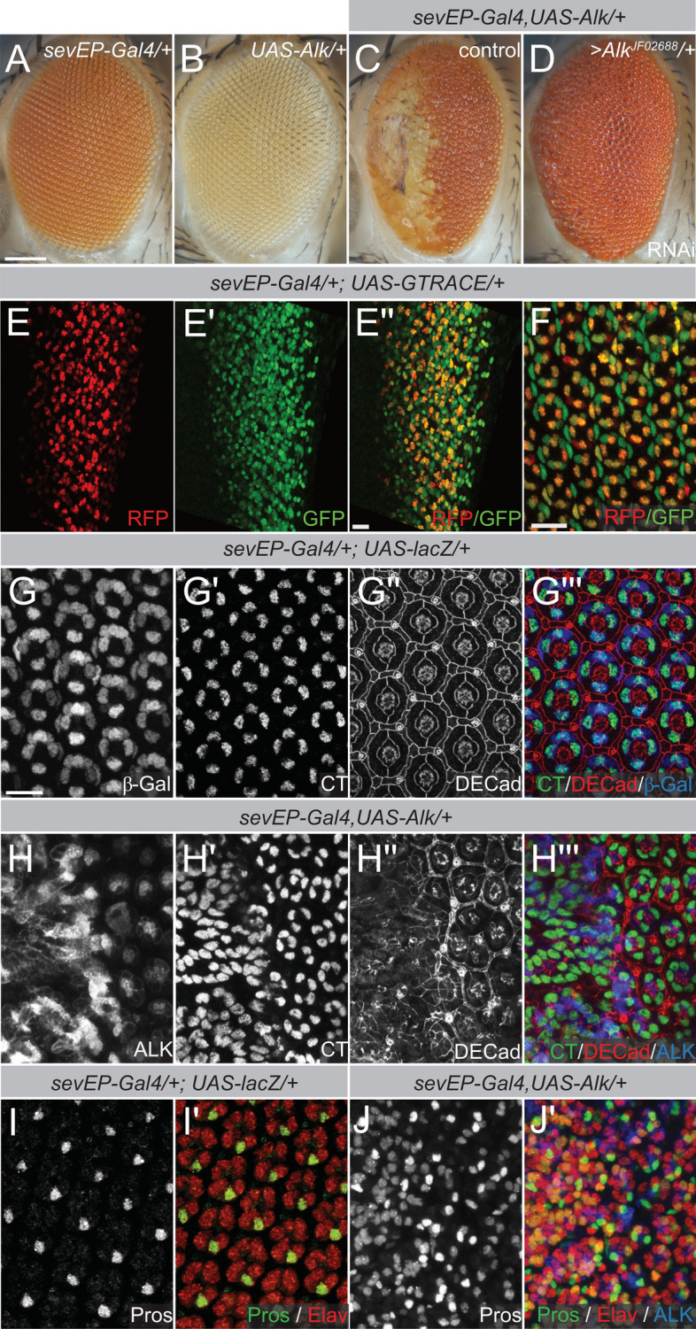
Ectopic expression of UAS-Alk/ + with the sevEP-Gal4 driver interferes with normal eye development. (A–D) Eyes of adult female flies with the indicated genotypes are shown. n > 200, 100% penetrant (A) “Wild-type” eye morphology of flies with either one copy of the sevEP-Gal4 driver insertion or (B) one copy of the UAS-Alk transgene. (C) Ommatidial disruption and necrotic scars in the anterior eye upon sevEP-Gal4/ + induced UAS-Alk/ + expression. The posterior half exhibits fused ommatidia and missing bristles. (D) Phenotypic rescue by co-expression of an Alk-specific RNAi. (E,F) G-TRACE analysis. Real-time sevEP-Gal4 expression is indicated by RFP (red) while continued expression in the lineage is indicated with GFP (green). (E–E’’) G-TRACE analysis in larval eye discs and in pupal eye discs (F). (G–J’) Immunofluorescence staining of eye discs from pupae (50 h apf) of the indicated genotypes. Antibody staining against Cut (CT) and Drosophila epithelial cadherin (DECad) reveals increased numbers of CT-positive cells and loss of ommatidia organization upon ectopic Alk expression (quantified in Fig. S1). (G,G’’’) βGalactosidase (βGal) reveals driver activity in a control eye disc, (H,H’’’) anti-ALK staining reveals transgene expression. Elav labels all photoreceptor cells and anti-Prospero (Pros) staining was used to identify R7 cells (I–J’). (J,J’) Upon ectopic expression of UAS-Alk the number of R7 photoreceptors is increased (quantified in Fig. S1). Scale bars are 100 µm in (A) and 10 µm in (E’’) and (F,G); anterior is left in all images.