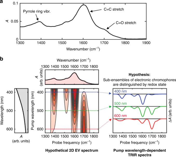
Fig. 3. Vibrational spectroscopy of DOPA melanin and UVF predictions.
a FTIR spectrum of DOPA melanin dissolved in phosphate-buffered D2O, including vibrational assignments. b Ultrafast vibrational fingerprinting (UVF) spectroscopy vs. two-dimensional electronic-vibrational (2D EV) spectroscopy for a hypothetical sample containing multiple electronic chromophores that can be distinguished by the redox state of their oxidizable IR-active functional groups. With time-resolved infrared (TRIR) spectroscopy (right), a subensemble of eumelanin chromophores is photoselected using a particular visible wavelength and its time-resolved vibrational spectrum is measured. These spectra are essentially horizontal slices through the hypothetical 2D EV spectrum (left). The 2D signals are assumed to only contain negative absorptive (bleaching) contributions for simplicity. Note that the presence of positive signal contributions from new vibrational frequencies in excited electronic states can alter the positions of the negative bands.

