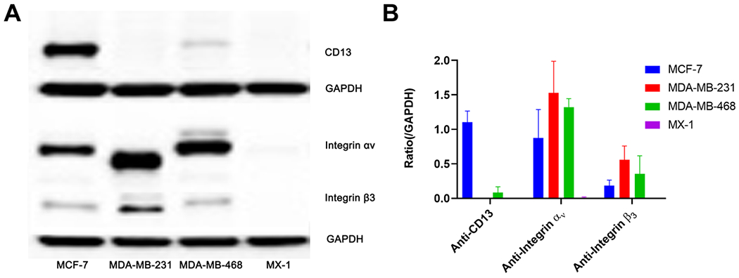
Figure 2.

(A) Western blotting was performed to assess the expression levels of CD13, integrin αv, and integrin β3 in MCF-7, MDA-MB-231, MDA-MB-468, and MX-1 cells with GAPDH used as the internal control. (B) The semi-quantitative analysis was conducted through the integrated optical density ratio of CD13, integrin αv, and integrin β3 to GADPH (data represent one of three separate experiments, mean ± SD).
