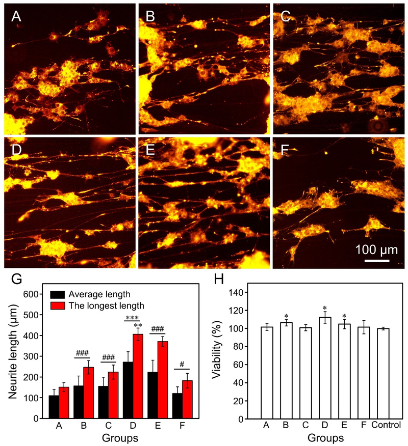
Figure 2.
(A−F) Fluorescence micrographs showing the neurite extending from PC12 cells after culture on the uniaxially aligned PCL microfibers (A) covered by a smooth surface, and decorated with nanoscale (B−E) grooves and (F) pores, respectively. Note that the microfibers used in Groups A to F corresponded to the same batches shown in Figure 1. (G) The average and longest lengths of neurites extending from the PC12 cells after culturing on the different types of microfibers. ***P < 0.001 as compared with that of cells cultured on all the other groups for the average length or that of cells cultured on all the other groups except Group E (**P < 0.01) for the longest length. ###P < 0.001 and #P < 0.05 as compared with that of cells cultured on PCL microfibers covered by a smooth surface. (H) Viability of PC12 cells after culturing on the different types of microfibers for three days. *P < 0.05 as compared with that of cells cultured on the control group (glass slides).

