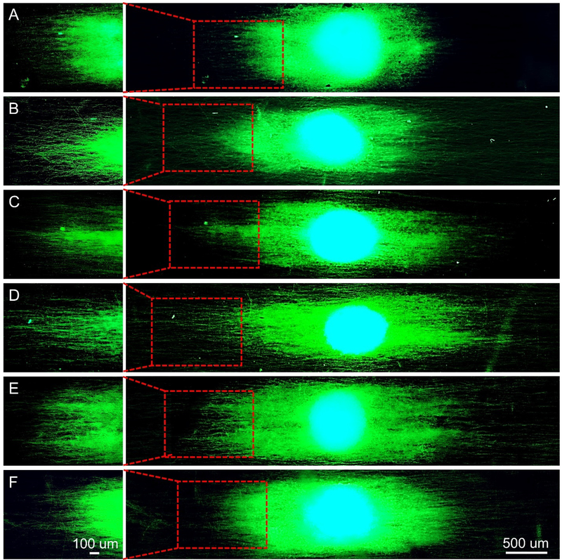
Figure 3.
Fluorescence micrographs showing typical neurite fields extending from DRG bodies when cultured on the uniaxially aligned PCL microfibers (A) covered by a smooth surface, and decorated with nanoscale (B−E) grooves and (F) pores, respectively. The neurites were stained with Tuj1 primary antibody (green) and cell nuclei were stained with 4′,6 diamidino-2-phenylindole (blue). Note that the microfibers used in Groups A to F corresponded to the same batches shown in Figure 1.

