
Fig. 1. Ribbon model of FAP structure.
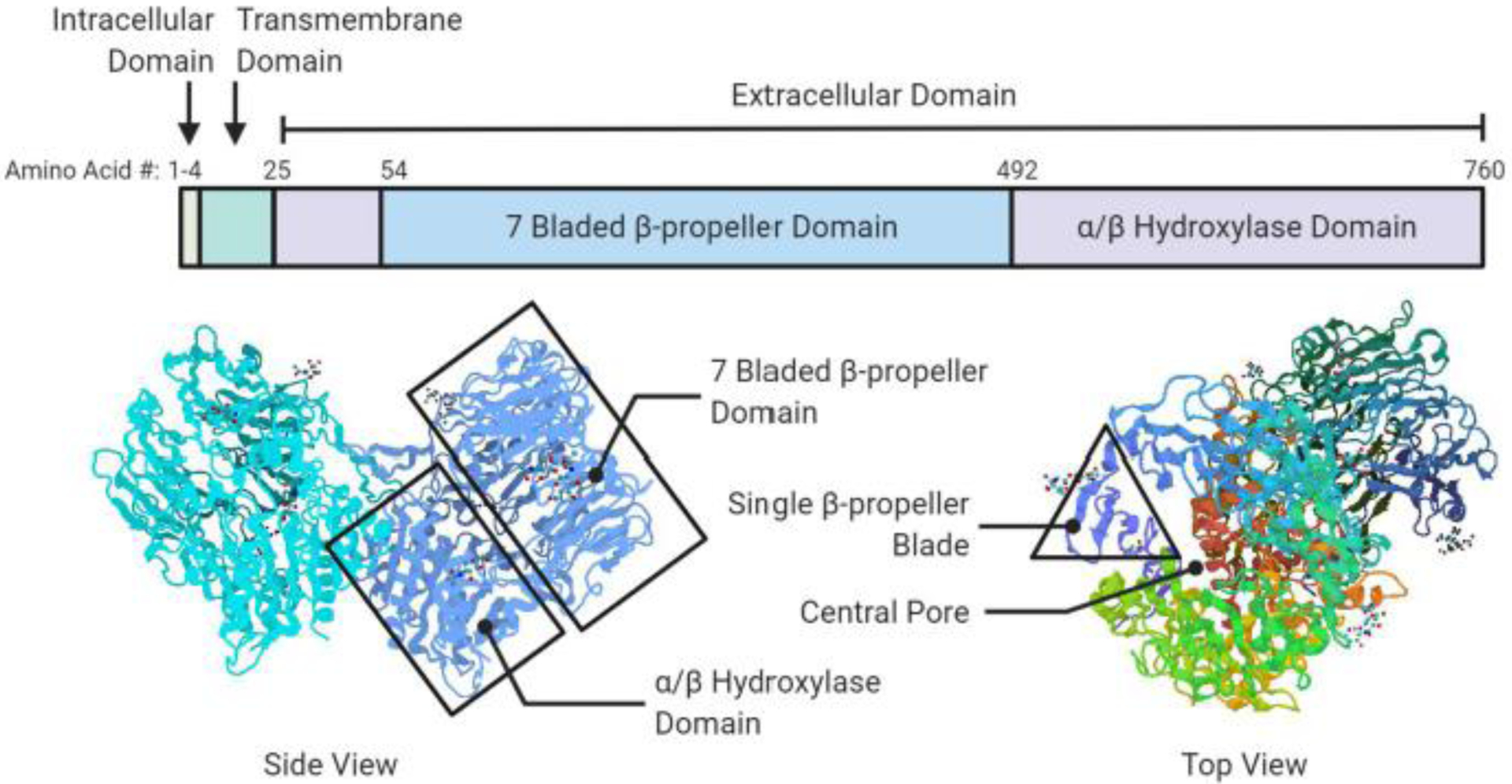
A schematic diagram of FAP domain structure (top) and ribbon models (bottom) depicting the FAP dimer. The seven-bladed β-propeller domain, αβ hydroxylase domain and β-propeller blade are highlighted. Figure created in biorender.com, PDB ID# 1Z68 [25].