
Figure 3.
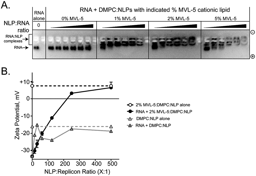
Cationic NLPs form complexes with replicon RNA. (A) Agarose gel electrophoresis demonstrates interaction between replicon RNA and cationic NLPs. In this representative example, apoA1 NLPs prepared with increasing percentages of MVL-5 (0, 1, 2, and 5%) were incubated with replicon RNA (NLP:RNA ratios = 15:1, 31:1, 62.5:1, 125:1, 250:1, and 500:1) for 30 minutes prior to agarose gel electrophoresis. Decreased RNA mobility was observed at higher NLP:RNA ratios. Non-cationic NLPs (0% MVL-5) had no effect on RNA mobility. Gels were stained with SybrGold to visualize RNA. (B) Zeta potential was characterized for representative NLP:replicon formulations. RNA complexes with cationic NLPs (circles, black trace; apoA1, DMPC, 2% MVL-5) trend toward increasingly positive zeta potential at increasing NLP:RNA ratios. Non-cationic NLPs (triangles, gray trace; apoA1, DMPC, 0% MVL-5) do not alter the intrinsically negative zeta potential of the RNA replicon. Images are representative of 3 independent experiments.