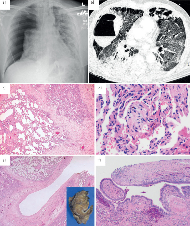
FIGURE 1.
Radiology and pathology in pneumothorax coronavirus disease 2019 (COVID-19). a) Anteroposterior erect chest radiograph: a male is his sixties presenting with a large right pneumothorax and some leftward tracheal shift. Background widespread bilateral alveolar opacity is consistent with “classic” COVID. b) Axial computed tomography image of the thorax acquired in a COVID-19 patient shortly before development of a right-sided pneumothorax. Note a large right-sided thin-walled cavity with air–fluid level, as well as numerous subpleural cystic spaces in the anterior hemithoraces bilaterally. c) Medium-power photomicrograph of lung parenchyma showing foci of collapse with accompanying fibrosis and vascular congestion. d) High-power image of intra-alveolar fibromyxoid plugs, fibrin and haemosiderin deposition. e) Low-power view of the 15-mm cystic space with a thick, fibrotic wall (inset: corresponding macroscopic cross-section). f) Medium-power image of the fibrous cyst wall (right) transitioning with respiratory epithelium (left), suggesting possible connection with the bronchial tree.

