
Figure 1.
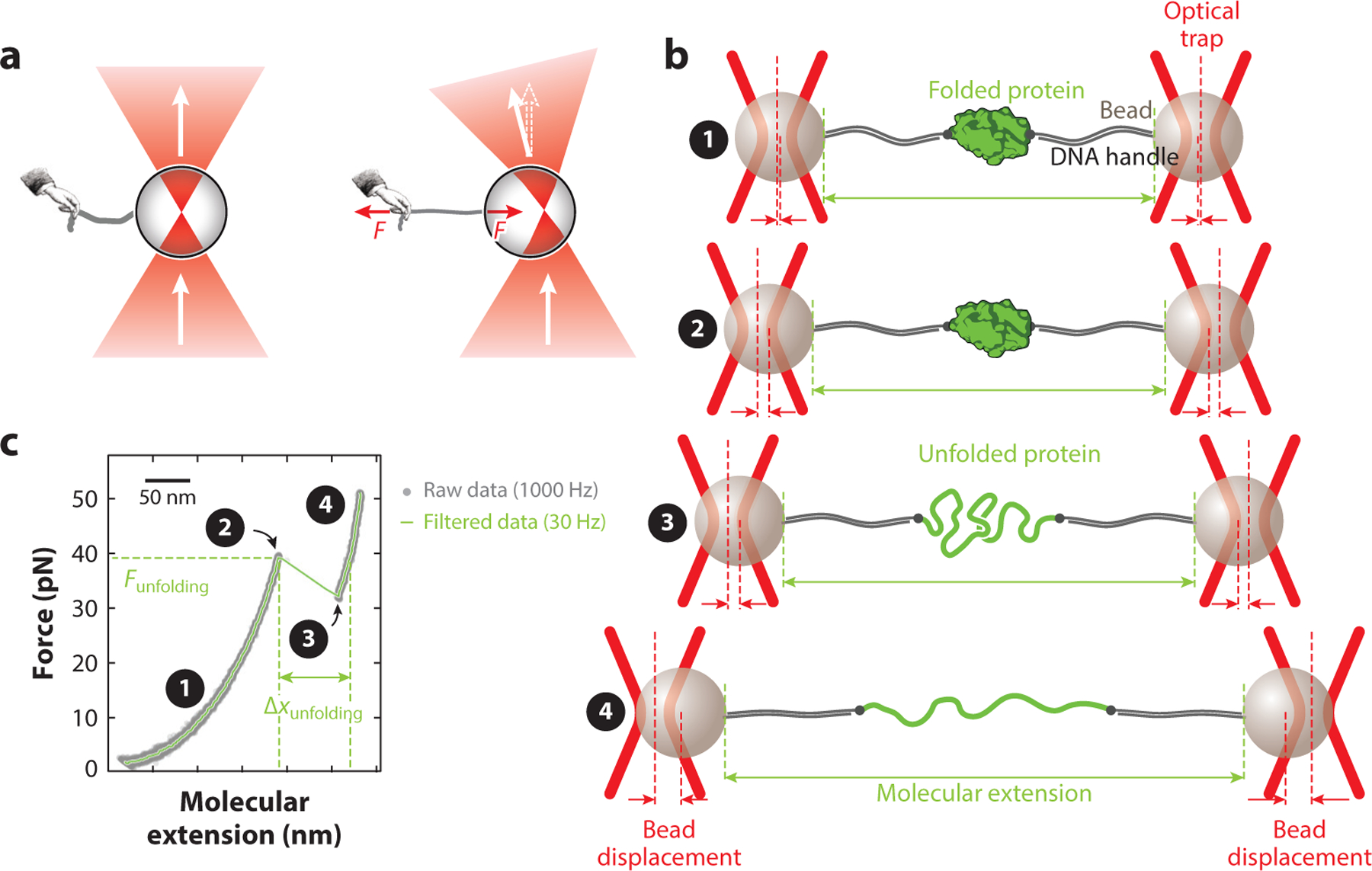
Manipulation of single molecules using optical tweezers. (a) Light from a tightly focused laser beam (red) passes through a bead. When the center of the bead is aligned with the laser focus (left), all rays hit the bead surface at a right angle, and the light does not change direction. When the center of the bead is not aligned with the laser focus (right), the change in momentum of the light elicits an equal and opposite change in momentum of the bead, resulting in a force F that attracts the bead to the laser focus. (b) Schematic of a protein tethered between two trapped beads via DNA handles (not drawn to scale). At low forces, the protein remains in the folded state (① and ②). However, an increase in force results in stretching of the DNA handles, increasing the molecular extension of the assembly between the beads. Unfolding of the protein results in a further increase in extension (③). Further increasing the force results in stretching of the DNA and the unfolded protein (④). Note that the bead displacement is proportional to the applied force, because the traps behave as harmonic springs. (c) Example of a typical force–extension curve, generated by applying a continuously increasing force to a tethered protein; gray dots represent data at 1,000 Hz, and the green curve represents data filtered to 30 Hz. The numbers are as in panel b. The curvature is due to the entropic elasticity of the DNA (region ①) and DNA plus unfolded protein (region ④). Unfolding of the tethered protein is apparent as a discontinuity in the curve (rip; from point ② to point ③).