FIGURE 3.
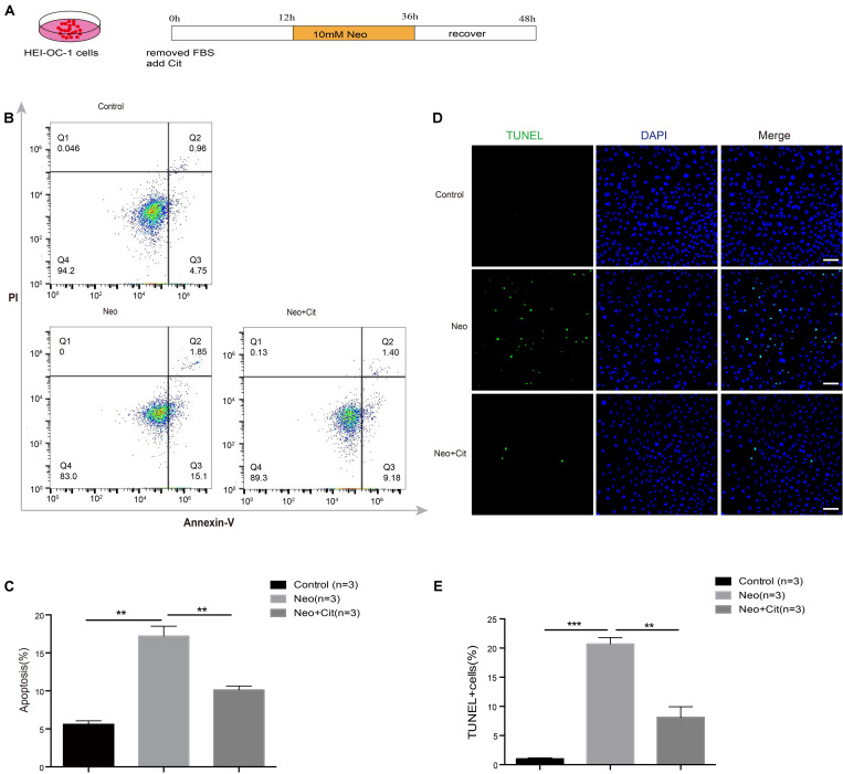
Citicoline protects against apoptosis in HEI-OC-1 cells after neomycin exposure. (A) Schematic diagram of citicoline (Cit) and neomycin addition in cell culture. (B) Apoptosis analysis by flow cytometry after different treatments. The upper right quadrants, lower right quadrants, and lower left quadrants of the images represent late apoptotic cells, early apoptotic cells, and live cells, respectively. (C) Flow cytometry results showing that the percentages of apoptotic cells after neomycin treatment were significantly higher compared with the undamaged controls. The amount of apoptosis induced by neomycin was significantly reduced by pretreatment with citicoline. (D) TUNEL and DAPI double staining showing the apoptotic HEI-OC-1 cells after different treatments. (E) The number of TUNEL/DAPI double-positive cells after neomycin exposure was significantly reduced by treatment with citicoline. Data are shown as mean ± SD. *p < 0.05, **p < 0.01, ***p < 0.001. Scale bars = 20 μm.

