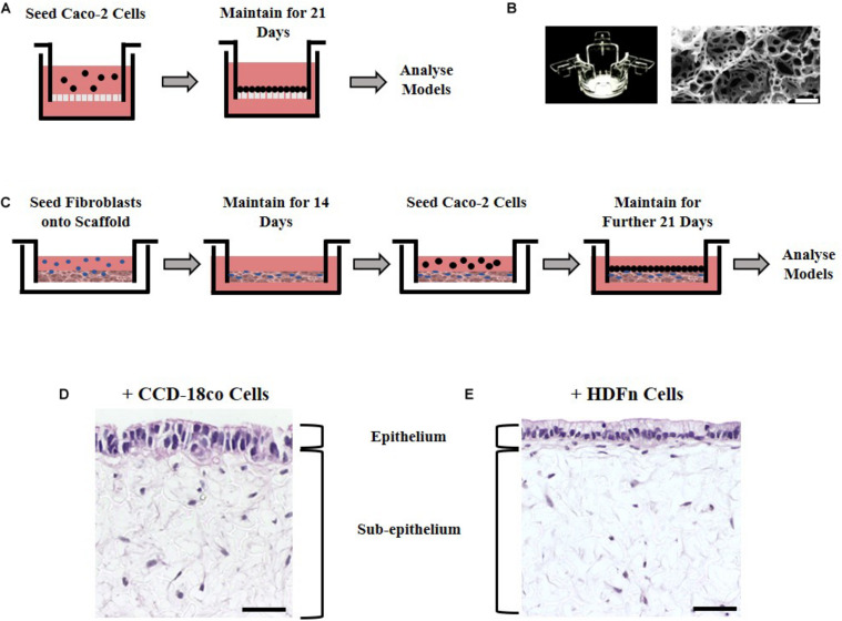
FIGURE 1.
Experimental set up in building models of the intestine. (A) Schematic representation of Caco-2 cell culture methodology on Transwell® inserts. (B) Alvetex® well insert and scanning electron micrographs showing the structure of the Alvetex® Scaffold membrane used in the co-culture models. (C) Schematic protocol depicting how fibroblast and epithelial cells were grown together in the 3D co-culture models. (D) H&E images of the resulting 3D co-culture models showing Caco-2 cells growing on top of Alvetex® Scaffold populated with either one of the two types of fibroblasts tested: (D) CCD-18co; (E) HDFn. Scale bars: 50 μm.

