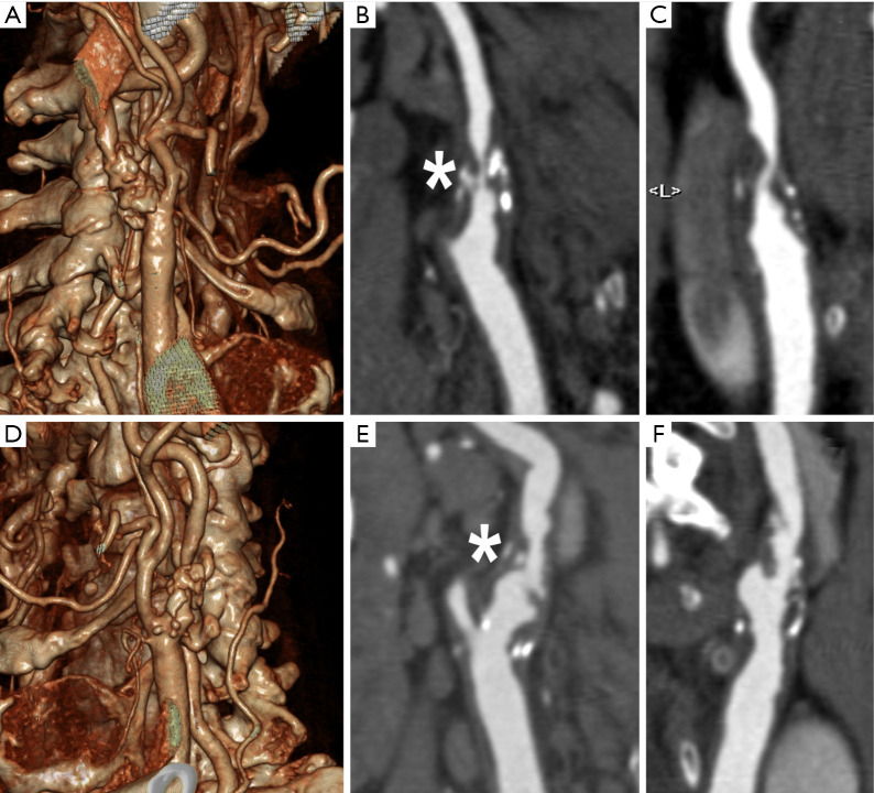
Figure 1.
Example of multislice CT Angiography of the Carotid arteries showing bilateral complex atherosclerotic plaques of the carotid bifurcation and internal carotid artery proximal segment. (A,B,C) Figures show the 3D Volume Rendering (A) and multiplanar reformats (B,C) of the right carotid artery characterized by a large plaque with mixed components (calcified and non-calcified) and concentric pattern, determining a high grade stenosis >70% (* in B); the plaque shows some very hypodense region suggestive for lipid content. (D,E,F) Figures show the 3D Volume Rendering (D) and multiplanar reformats (E,F) of the left carotid artery characterized by a large plaque with mixed components, but most of all the intimal surface appears ulcerated, and even though the degree of stenosis is lower on this side this plaque has a higher risk (* in E).

