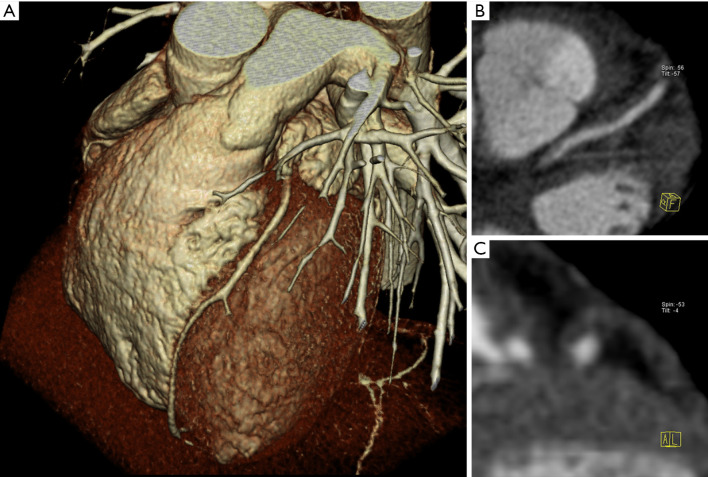
Figure 2.
Example of multislice CT Angiography of the coronary arteries. On 3D Volume Rendering the left anterior descending coronary artery appears normal without any significant stenosis (A). However, multiplanar reformats (B,C) clearly show a long and completely non-calcified plaque with positive remodeling. This pattern represents a high risk plaque in the coronary artery tree.

