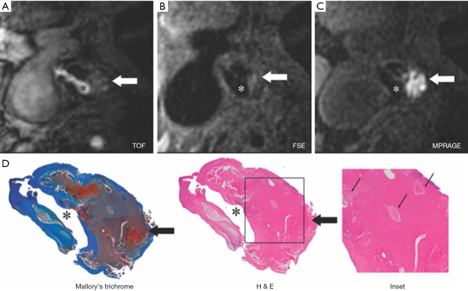
Figure 3.
Moderately calcified IPH in left internal carotid artery in 75-year-old man. (A,B,C) On T1-weighted MR images, high-signal-intensity area (arrow) measures 15.8 mm2 on the TOF image, 8.6 mm2 on FSE image, and 24.8 mm2 on MPRAGE image; (D) mallory trichrome–stained histologic specimen shows variable red staining pattern in necrotic core (arrow) (Original magnification, ×10). Adjacent section stained with hematoxylin-eosin (H&E) shows large necrotic core (arrow) filled with IPH measuring 34.9 mm2. (Original magnification, ×10.) The inset is a magnified region of necrotic core (outlined in black) showing small fragments of calcification (arrows). (Original magnification, ×100.) *, the lumen of the left internal carotid artery. TOF, time-of-flight MR angiography; FSE, fast spin-echo; MPRAGE, magnetization-prepared rapid gradient echo; IPH, intraplaque hemorrhage. (From Ota H, Yarnykh VL, Ferguson MS, et al. Carotid Intraplaque Hemorrhage Imaging at 3.0 T MR Imaging: Comparison of the Diagnostic Performance of Three T1-weighted Sequences. Radiology 2010;254:551-63 with permission.)

