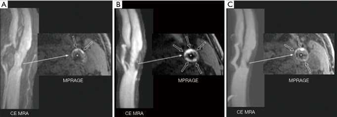
Figure 4.
Presence of IPH is associated with plaque growth and increasing stenosis on standard statin therapy that regressed with intensive statin therapy as seen on serial research carotid plaque MR studies. (A) This 76-year-old asymptomatic patient who had been on atorvastatin 10 mg for 3 years presented with a 51% stenosis of the left internal carotid artery on CE MRA. On 3D MPRAGE images, IPH was seen as the focally intense region (open arrows); (B) despite increased statin therapy with atorvastatin 40 mg for 2 years, there was a progression in the size of the IPH (open arrows) and increase in carotid stenosis to 69%; (C) based on the plaque and stenosis progression, statin therapy was increased again to 80 mg of atorvastatin. Over the next 2 to 3 years there was clear regression of the IPH (open arrows) with the carotid stenosis now measuring 58%. *, internal carotid artery lumen. IPH, intraplaque hemorrhage; CE MRA, contrast-enhanced MR angiography; MPRAGE, magnetization-prepared rapid gradient echo. (From DeMarco JK, Spence JD. Plaque Assessment in the Management of Patients with Asymptomatic Carotid Stenosis. Neuroimaging Clin N Am 2016;26:111-27 with permission.)

