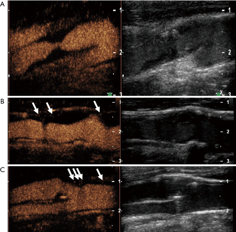
Figure 4.
Semi-quantitative, visual-based analysis of intraplaque neovascularization using a 3-point grading system. Plaques at the origin of the internal carotid artery on B-mode ultrasound (right side) and CEUS imaging (left side) in three different patients. Grade 1 (A): carotid plaque with no intraplaque neovascularization defined as no appearance of moving microbubbles in the plaque or confined only to the adjacent adventitial layer. Grade 2 (B): carotid plaque with limited or moderate intraplaque neovascularization defined as moderate visible appearance of moving bubbles in the plaque at the adventitial side or plaque shoulder (arrows). Grade 3 (C): carotid plaque with extensive intraplaque neovascularization defined as clear visible appearance of bubbles moving to the plaque core (arrows).

