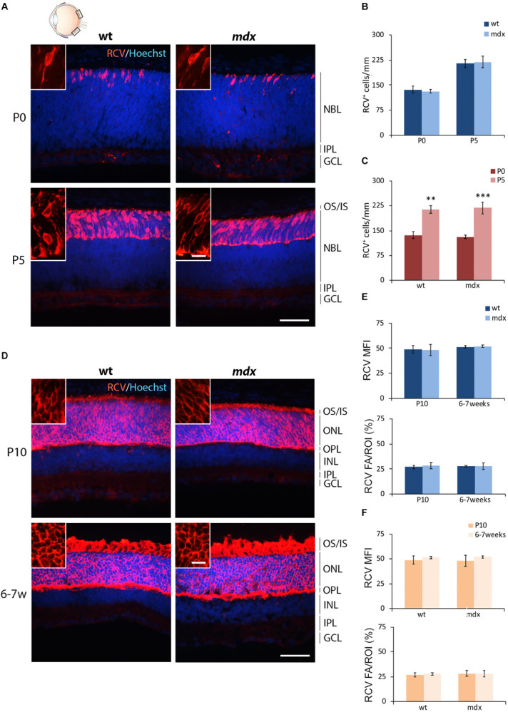
FIGURE 2.

Lack of Dp427 does not alter neither the number of recoverin-immunopositive photoreceptors nor the recoverin fluorescence intensity. (A,D) Representative images of horizontal (N–T) retina cryosections of P0, P5, P10, and 6–7-week-old wild type (wt) and mdx mice immunolabeled for recoverin (RCV), a pan-photoreceptor cell marker. Nuclei are stained in blue (Hoechst). (B,C) Comparative quantitative analysis of the number of RCV+ cells/mm of retina between wt and mdx age-matched mice (B) and across post-natal days within the same genotype (C). (E,F) Comparative quantitative analysis between wt and mdx age-matched mice (E) and across post-natal days within the same genotype (F) based on both mean fluorescent intensity (MFI) (normalized against the background) and percentage of RCV fluorescent area (FA)/ROI area, measured by the ImageJ software. Both cell counts (B,C) and measures of the fluorescence intensity (E,F) are made in the posterior retina (black boxes in the eye drawing). Data are analyzed by two-way ANOVA test Sidak post hoc and represented as the mean ± SEM. ∗∗p ≤ 0.01, ∗∗∗p ≤ 0.001 (P5 vs. P0 in C). n = 3–5 independent experiments. Scale bar: 50 μm; insets: 10 μm. OS, outer segment of photoreceptors; IS, inner segment of photoreceptors; NBL, neuroblastic cell layer; ONL, outer nuclear layer; OPL, outer plexiform layer; INL, inner nuclear layer; IPL, inner plexiform layer; GCL, ganglion cell layer.
