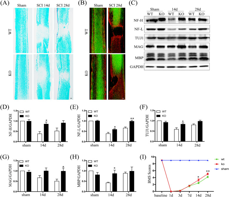
Fig. 8.
Hv1 deficiency promotes axonal regeneration and recovery of motor function. a Representative image of Luxol fast-blue staining on days 14 and 28 after SCI (scale bar, 200 μm). b Representative image of GFAP staining (red) and BDA-labeled axons (green) in sham-operated mice and at 28 days after SCI in WT and KO mice (scale bar, 200 μm). c Western blotting showing NF-H, NF-L, TUJ1, MAG, and MBP levels in sham-operated mice and at 14 and 28 days after SCI. d–h Quantification of Western-blot results for NF-H (d), NF-L (e), TUJ1 (f), MAG (g), and MBP (h) normalized to GAPDH. Values are represented as the mean ± SEM (n = 5 for each treatment). i Analysis of Basso Mouse Scale (BMS) scores before and after SCI in WT and KO mice (n = 8 for each genotype; *p < 0.05 **p < 0.01, KO SCI vs. WT SCI)

