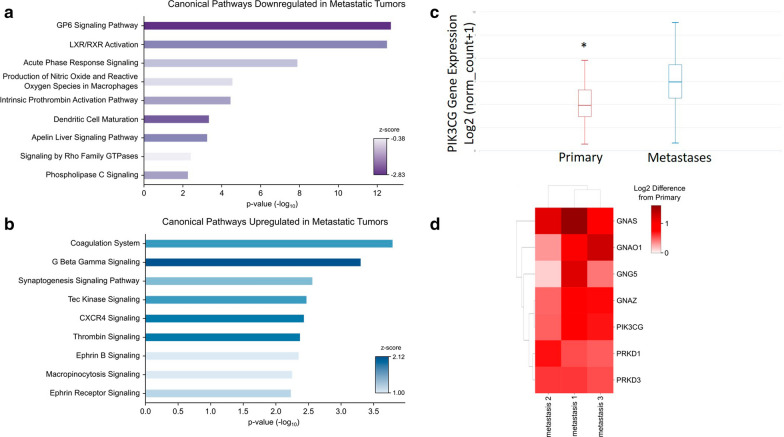
Fig. 3.
Pathway Analysis identifies PI3KCG as upregulated in MBM tissue. a Canonical pathways downregulated in the metastatic brain tumors. b Canonical pathways upregulated in the metastatic brain tumors. c PIK3CG expression in the cancer genome atlas melanoma cohort demonstrates significantly decreased expression in primary tumor tissue in comparison to metastatic tissue (p = 4.77 × 10⁻¹⁵). d Heatmap of proteins in common among the G beta gamma, CXCR4, and thrombin signaling pathways significantly increased levels of PI3KCG protein in the MBMs compared to the primary tumor

