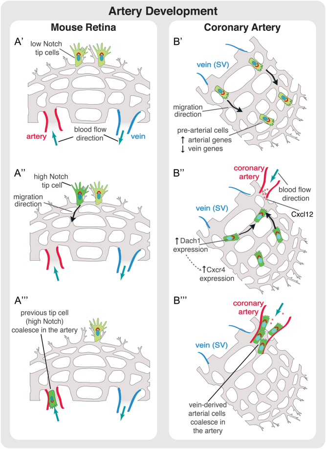
Figure 3.
Cell migration during artery development. (A) Proposed model of artery development during angiogenesis in the mouse retina. (A’) Tip cells are activated at the sprouting front by VEGF signaling. (A‘‘) Some tip cells are activated by Notch signaling that promotes their migration away from the sprouting front in direction of the artery. (A‘‘‘) These previous tip cells coalesce in the artery leading to artery growth. (B) Proposed model for coronary artery development. (B’) Vein ECs derived from sinus venosus (SV) sprout by angiogenesis. (B‘‘ During this stage, and prior to the onset of blood flow, some SV-derived ECs start to increase the expression of arterial markers reducing the expression of venous ones. This genetic profile modification, accompanied to the onset of blood flow, induces migration of these pre-arterial cells. The migration of EC against flow direction is regulated by the transcription factor DACH1. DACH1 stimulates expression of CXCR4 in pre-arterial cells enabling these cells to respond to CXCL12, which is expressed by arterial cells. (B‘‘‘) ECs connect to the coronary artery leading to its growth and development.

 This work is licensed under a