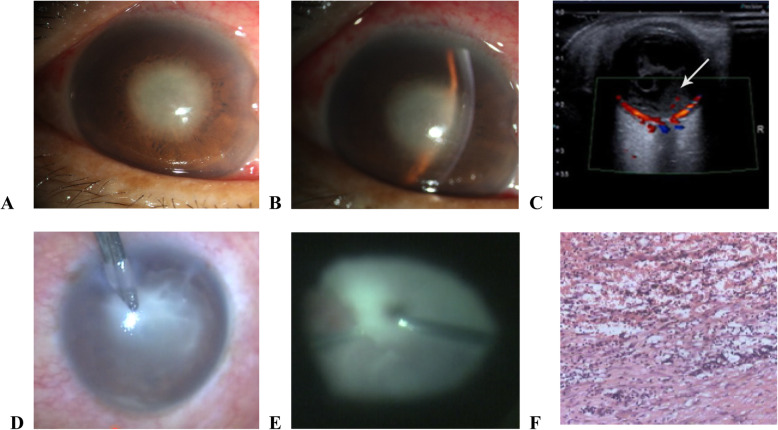
Fig. 2.
Clinical features of liver endophthalmitis. a and b Biomicroscopic examination revealing conjunctival hyperemia, corneal edema, and inflammation of the anterior chamber, accompanied by discernable pus accumulation or fibrinous exudates. c Three-dimensional ultrasound imaging of the eye showing turbidity in the form of streaks, dots, or floccules in the vitreous humor. d-e Purulent exudate was observed in the anterior chamber and vitreous humor. Tight adhesion of fibrinous exudate was observed in the vitreous cavity. f Pathological image of eye biopsy showing extensive neutrophil infiltration and piecemeal necrosis

