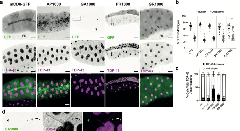
Fig. 4.
TDP-43/TBPH mislocalisation in DPR expressing Drosophila. a TDP-43/TBPH (magenta) and DPR (green) localisation in Drosophila Salivary glands. Scale bars 50 μm boxed region expanded in (d). b Quantification of the percentage of TDP-43/TBPH localising to the nucleus or cytoplasm (60 cells from 3 animals, from 3 independent crosses, per genotype). c Quantification of the percentage of DPR containing cells with insoluble TDP-43/TBPH inclusions. Total number of DPR containing cells (n) are shown on bars, total number of animals (N) = 3 per genotype. d co-localisation of TDP-43/TBPH and PolyGA aggregates

