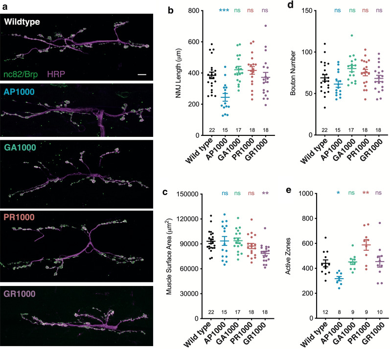
Fig. 6.
Morphological analysis of the Drosophila larval neuromuscular junction. a Micrographs showing the neuromuscular junction (NMJ) (muscle 6/7 hemi-segment A3) of third instar larvae pan-neuronally expressing (nSyb-Gal4) DPRs. Anti-HRP labels the nervous system (magenta) and anti-bruchpilot (Brp/nc82) active zones (green). Scale bars 10 μm. Quantification of b NMJ length, c muscle surface area, d bouton number and e active zone number. ANOVA with post hoc Dunnett’s multiple comparison to wild type controls ***p < .001; **p < .01; *p < .05. The number of NMJ’s analysed are shown on each graph. NMJs were quantified from at least 8 animals (N = 8) taken from at least 3 independent crosses per genotype

