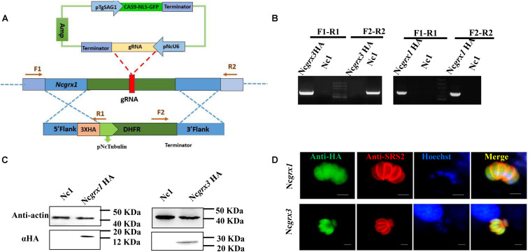
FIGURE 2.
The cellular localization of NcGrxs. (A) Strategy for construction of the NcGrx1-HA and NcGrx3-HA parasites. (B) The insertion of 3 × HA-tagged NcGrxs in parasites was confirmed by PCR. (C) Western blot confirmed the expression of HA-tagged NcGrxs in parasites. Actin was used as a control. (D) Immunofluorescence assays analysis of NcGrxs localization. αHA was used to detect the NcGrxs-HA (green), and SRS2 (red) served as a parasite surface marker (bar = 5 μm).

