Fig. 6.
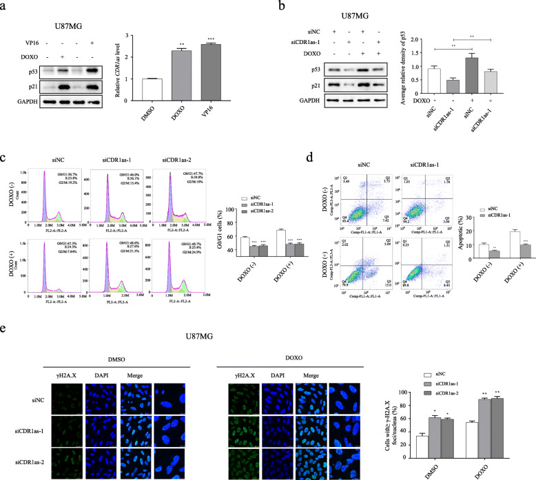
CDR1as serves as a protective machinery to preserve p53 function against DNA damage in U87MG cells. a Western blot analysis of p53 and p21 expression (left); and RT-qPCR analysis of CDR1as expression (right) in U87MG cells after 48 h treatment with DOXO, VP16 or DMSO (as control). b Western blot analysis of p53 and p21 expression (left); and densitometric analysis of p53 expression normalized to GAPDH (right) in U87MG cells transfected with siCDR1as-1 or siNC after 48 h treatment of DOXO or DMSO. c Flow cytometric analysis of cell cycle in U87MG cells transfected with different siCDR1as or siNC after 48 h treatment of DOXO or DMSO. d Flow cytometric analysis of apoptosis in U87MG cells transfected with siCDR1as-1 or siNC after 48 h treatment of DOXO or DMSO. e IF analysis of γH2A.X (DNA damage marker) in U87MG cells transfected with different siCDR1as or siNC after 48 h treatment of DOXO or DMSO (left); quantification of number of γH2A.X positive cells with equal or more than 10 γH2A.X foci/nucleus (right). *p < 0.05; ** p < 0.01; ***p < 0.001

