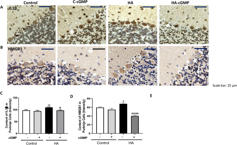
Fig. 10.
Extracellular cGMP normalizes the IL1β and HMGB1 in Purkinje cells of hyperammonemic rats. Immunohistochemistry was performed as indicated in methods with DAB staining using antibodies against IL1β (a) and HMGB1 (b). Representative images are shown. The content of IL1β (c) and HMGB1 (d) was quantified in Purkinje cells as described in the “Methods” section. Values are mean ± SEM of 6 rats per group. Values significantly different from control rats are indicated by asterisk and from hyperammonemic rats are indicated by “a”. *p < 0.05, **p < 0.01; a p < 0.05, aaaa p < 0.0001. Data were analyzed using a one-way analysis of variance (ANOVA) followed by Turkey post hoc

