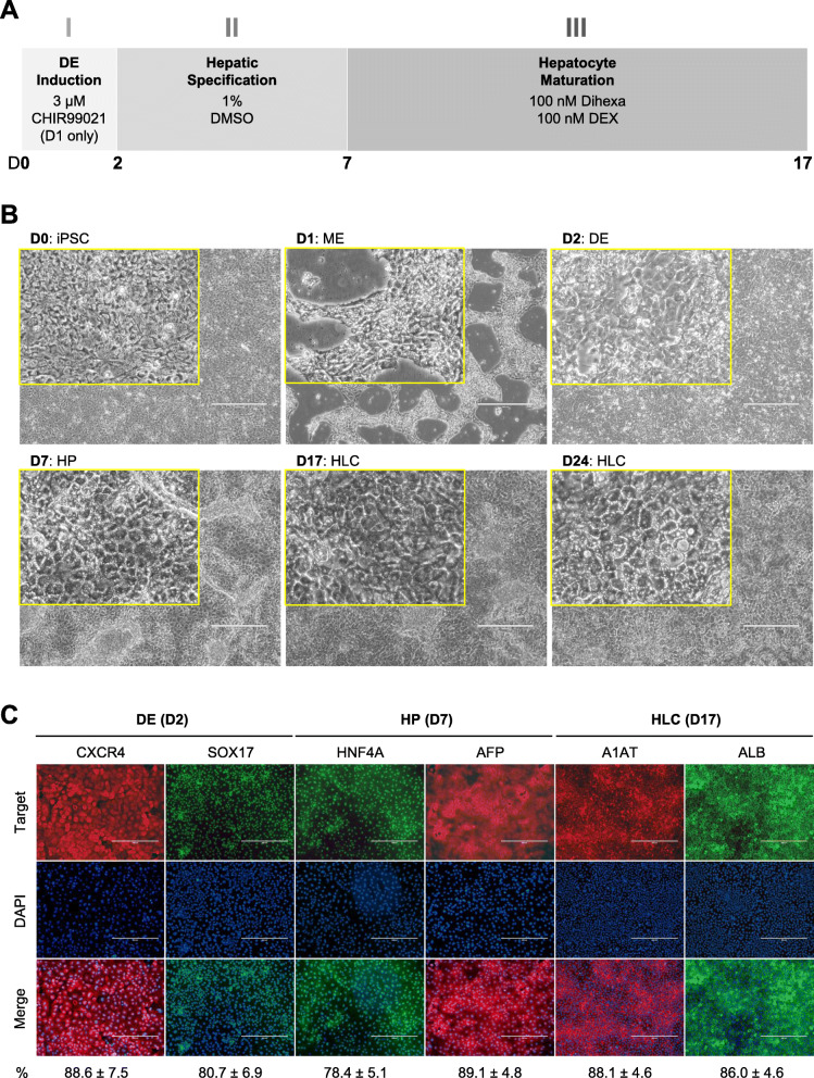
Fig. 1.
Small molecule-based protocol for the differentiation of hepatocytes from induced pluripotent stem cells. a Schematic diagram showing the three-phase differentiation process. b Representative images showing sequential morphological changes at each stage of the differentiation. iPSC, induced pluripotent stem cell; ME, mesendoderm; DE, definitive endoderm; HP, hepatic progenitor; HLC, hepatocyte-like cell. Magnification: × 10, Insert: × 40, Scale bar: 400 μm. c Representative immunocytochemistry images demonstrating the expression of key markers at each stage of the differentiation. The percentage of marker positive cells, presented as mean ± SD of three independent experiments, is listed at the bottom under each marker. Scale bar: 400 μm

