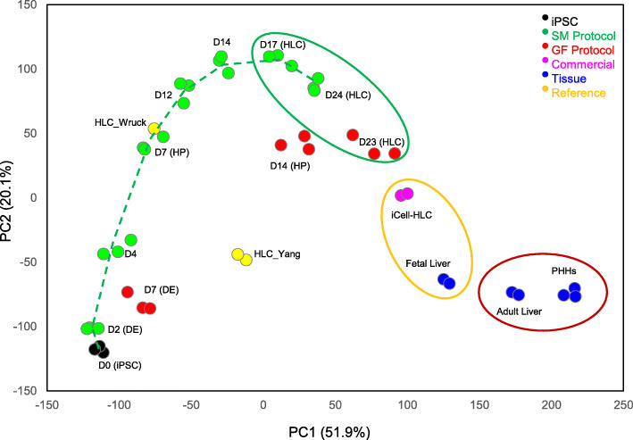
Fig. 3.
Principal component analysis (PCA) plot showing the differentiation track (dotted line) and similarities/dissimilarities among the samples. The two axes PC1 and PC2 represent the first two principal components identified by the analysis. The percentage contribution of each component to the overall source of variation is included in the parentheses following the component name. Color codes of different cell types are shown at the top-right corner. The timing of the small molecule-driven and growth factor-driven differentiation is denoted by D (day) followed by the number of the day. iPSC, induced pluripotent stem cell; DE, definitive endoderm; HP, hepatic progenitor; HLC, hepatocyte-like cell; PHHs, primary human hepatocytes. HLC_Wruck and HLC_Yang are reference datasets

