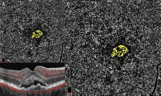
Figure 1.
(A) The 6 × 6 mm en face CC angiogram illustrates subfoveal treatment-naïve MNV (highlighted in yellow). (B) OCT B-scan shows the corresponding CC segmentation. The Bruch's membrane offset was used to segment the CC slab, defined as a 10-µm thick slab with an inner and an outer boundary located, 21 µm and 31 µm, respectively, below the anatomic location of the Bruch's membrane. (C) The perilesional DH is encircled in yellow and is represented by a low-flow area surrounding the MNV.

