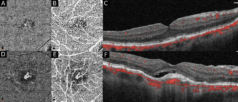
Figure 4.
A subanalysis was carried out to assess the differences in the percentage (%) of CC FD in exudative versus nonexudative treatment-naïve MNV. Compensated en face CC OCTA slabs (A, D) are binarized using the Phansalkar local thresholding method (shown here) on Image J. (The standard deviation global thresholding method was also performed in this analysis to validate the Phansalkar method.) The CC FD is quantified after masking the superficial and large vessels (D, E). The B-scans (C, F) are used to determine the nature of the MNV, nonexudative (C) versus exudative (F).

