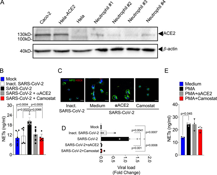
Figure 4.
SARS-CoV-2 infection in neutrophils depends on ACE2 and serine protease TMPRSS2 pathway for the NETs formation. (A) Expression of ACE2 was assessed by Western blot (A) in Caco-2, HeLa cells transduced with hACE2 (Hela-ACE2), HeLa cells, and isolated neutrophils from healthy controls. β-Actin expression was used as load control for protein expression. SARS-CoV-2–infected neutrophils (MOI = 1.0) were pretreated with neutralizing anti-ACE2 antibody (αACE2, 0.5 µg/ml) and camostat (10 µM), a serine protease TMPRSS2 inhibitor. (B) NETs quantification by MPO-DNA PicoGreen assay in these neutrophils supernatants (n = 6). (C) Immunostaining for nuclei (DAPI, blue), MPO (green), and H3Cit (red). Scale bar indicates 50 µm. (D) SARS-CoV-2 viral load detection in neutrophil cell pellet (n = 3) by RT-PCR 4 h after infection. Fold change relative to SARS-CoV-2 group. (E) PMA-stimulated neutrophils from healthy controls (n = 3) were pretreated or not with 0.5 µg/ml αACE2 and 10 µM camostat. NET quantification was assessed by MPO-DNA PicoGreen assay in neutrophils supernatants after 4 h of PMA stimulation. Data are representative of at least two independent experiments and are shown as mean ± SEM. P values were determined by one-way ANOVA followed by Bonferroni’s post hoc test (B, D, and E).

