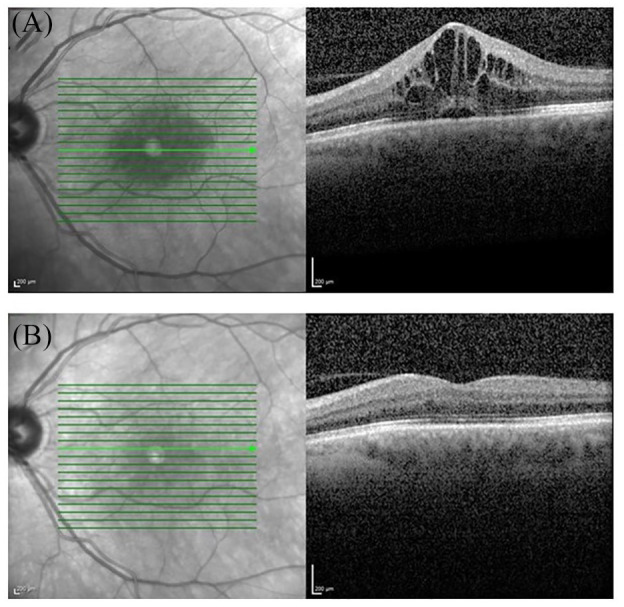
Figure 4.

Optical coherence tomography (OCT) in uveitic macula edema. (A) Cystoid macular edema and serous retinal detachment on a cross-sectional macular OCT scan. (B) Resolution of cystoid spaces and subfoveal fluid following treatment. Vision improved from 0.5 (A) to 1.0 (B). OCT is a non-contact non-invasive technique that allows in vivo imaging of the retina, choroid, optic nerve head, retinal nerve fiber layer and the anterior structures of the eye.
